What Does Dense Breast Tissue Mean For Mammography?
Depending on the nature of the breast tissue, the result of the mammography is not available
The breast tissue of a woman is not the same and changed the course of life.
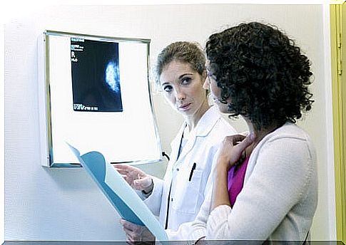
The significance of the mammography also depends on the nature of the breast tissue. The tighter the breast, the more difficult it is to recognize something in the recordings and to make reliable statements.
Differences in breast tissue
The breast tissue of a woman is not always the same, it is different for every woman and also changes in the course of life.
Breast tissue is a changeable mixture of fat, glandular tissue and connective tissue. The more connective tissue and the less fat there is in the breast, the denser the breast tissue.
The tighter the chest, the harder it is to see changes.
The type of breast tissue is genetically determined, a woman has a certain type of breast. There are four different types of breasts that differ in their density.
- ACR1: “fat transparent”; well transparent and easy to assess with mammography
- ACR2: moderately transparent on mammography
- ACR3: heterogeneously dense, decreased sensitivity in mammography
- ACR4: extremely dense, cancer and other changes in breast tissue not always detectable on mammography
Statistically, however, only about 4% of all women have very fat breast tissue (ACR1) and around 25% of women have very dense tissue. The vast majority of women are somewhere in between. The density decreases somewhat with age, but the basic breast type is retained.
What does this mean for mammography?
Depending on which device your doctor uses, there is a 2-D or 3-D mammography.
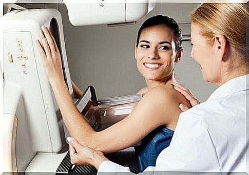
In the classic and most widely used mammography, a two-dimensional image of your breast is created using a special X-ray machine.
Modern devices can already create a three-dimensional image that makes the diagnosis much more reliable for the doctor, since he no longer has the problem of correctly recognizing and interpreting overlapping layers of tissue.
In order to obtain the X-ray image, each breast is placed individually in a special device and pressed between two plates with light pressure.
If the breast tissue is too dense, the combination of mammography and ultrasound delivers the best results, but ultrasound is an IGEL service in Germany, so it is not paid for by health insurance companies even with extremely dense breasts.
If the risk of breast cancer is high due to family history, you should seriously consider this additional examination as a supplement to mammography.
In principle, however, the following applies to all women, regardless of breast tissue: Mammography should never be used for diagnosis alone and should always be used in combination with other diagnostic methods.
Changes in the breast tissue can be made visible with mammography, but it cannot be clarified whether this change is benign or pathological.
This can only be clarified through other, further investigation methods. So if you’ve had a mammogram and it’s found changes, that doesn’t immediately mean you have breast cancer!
When should I have a mammogram?
The World Health Organization recommends mammography for the early detection of breast cancer.
She bases her recommendation on many significant international studies that show that mammography really does more benefit than harm.
Whether you ultimately use the free breast cancer screening options is up to you.
Just know that breast cancer is the deadliest cancer in women! In breast cancer, specialists always point out that detecting the disease early is the best way to cure it and extend life expectancy.
A mammography screening program has been offered in Germany for all women over 50 since 2005.
Women between the ages of 50 and 69 automatically receive an invitation by post every two years to have their breasts examined at specially trained mammography centers.
The data come from the residents’ registration office and the costs for the screening examination are covered in full by the statutory health insurance companies.
Women under 50 years of age do not receive an invitation, as the risk of a misdiagnosis increases with the age of the woman.
The younger the breast, the denser the breast tissue and the greater the chance of a wrong diagnosis.
In rural regions where there is no special mammography center, articulated trucks with trailers with a mobile mammography center are available on certain dates.
Is Mammography Harmful?
The radiation exposure during mammography is very low, since softer rays are used to image the soft tissue than when x-raying bones.
Nonetheless, the body is exposed to a certain amount of radiation. The radiation exposure on a long-haul flight on vacation is significantly higher than that of a mammography!