Peripatellar Pain Syndrome, Or Chondropathia Patellae: Features And Treatment
Peripatellar Pain Syndrome is a condition that is relatively common in young adults or adolescents who play sports. But older people who suffer from a degenerative disease such as osteoarthritis can also be affected.
A peripatelläres pain syndrome or chondromalacia patella is one of the most common knee disorders. Since this joint bears most of the human body weight, it is generally very prone to injury. In addition, there are other risk factors such as age, obesity, level of activity, etc.
First of all, you should know that peripatellar pain syndrome is a relatively common condition in young adults. Additionally, there is a higher incidence among those who practice the following sports: soccer, basketball, volleyball, tennis, cycling, karate, rowing, rugby, athletics, and ballet. Mountaineers are also very susceptible to chondropathia patellae.
There is some debate about the use of the name chondropathia patellae. The first term describes a softening of the cartilage and the patella is the kneecap. However, at some point it began to be used to diagnose anyone with knee pain.
Today , the scientific community believes that it would be more precise to speak of patellofemoral syndrome rather than peripatellar pain syndrome when the exact cause of knee pain is unknown. Still, the term is medically acceptable.
What is Peripatellar Pain Syndrome?
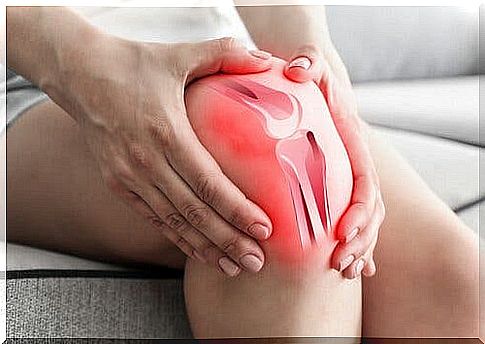
The kneecaps (patellae) are flat bones that are located at the front of the knee. They articulate the femur and their main function is to protect the joint while sliding and to facilitate this movement. Peripatellar pain syndrome is a degenerative condition that affects the cartilage on the articular surface of the kneecaps.
In this condition, the bone is exposed to direct friction due to joint movement. In most cases, this causes the cartilage to soften, causing pain on the front side of the knee.
The cartilage acts as a kind of shock absorber in the joints. It consists of around 90% water and 10% cells. As a result, it is able to withstand the energy of shocks. If patellar chondromalacia (a peripatellar pain syndrome) occurs, it is because the structure of the cartilage has changed. And then it is no longer smooth, but becomes rough and gray.
In addition, the cartilage becomes thinner and more irregular. Sometimes it cracks and even breaks. In these cases, you may hear a click or similar noise in your knee when you move it. In addition, there is also severe pain.
Causes of discomfort
The main cause of peripatellar pain syndrome is repeated trauma to the knee. In other words, repeated movements create compression in the cartilage. For this reason, athletes are also at an increased risk of this disease.
Likewise, the problem could be due to other factors, such as the following:
- Anatomical problems due to misalignment of the knee or improper placement of the kneecap
- Heavy blows to the knee. For example, if you fell on your bent knee or hit an object with your knee
- Inadequate walking due to an abnormality in your feet or legs, or from wearing high heels
- A previous knee dislocation or fracture
- Obesity
- Muscle wasting or weakness
- Legs of different lengths
- A pronounced curvature of the spine
Peripatellar pain syndrome: features
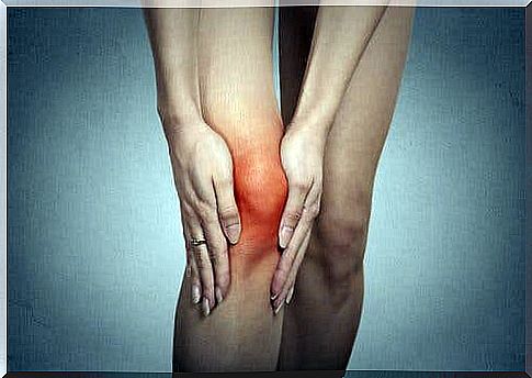
Typically, the pain occurs with physical activity. When the patient walks on hard ground or when walking up and down the stairs, these tend to be aggravated. Also, if you spend time in a position that requires keeping your knees bent for a long period of time, you may experience pain. In addition, stiffness also occurs in this case.
When the knee is bent, the characteristic cracking noises of the peripatellar pain syndrome often occur. Over time, this abnormality affects the strength of the leg muscles and makes walking difficult.
There are different degrees of severity depending on how this disease develops. These are:
- Grade 1. The cartilage softens and edema occurs.
- Grade 2. The diagnostic images show that the cartilage is fibrillating and fraying.
- Grade 3. The cartilage shows cracks, some of which are pronounced and extend into the deepest layers.
- Grade 4. Ulceration occurs. The 3rd degree condition worsens and the cracks are more noticeable and deeper.
- Finally, there is grade 5. This is where embolism occurs, that is, a pathological increase in cartilage density – a consequence of worsening ulceration that eventually attacks the subchondral bone.
In order to get this condition under control, conservative treatment is usually carried out first. If this does not work, however, an operation is required.